apical lung hernia
Medical College of Georgia. My attention was drawn to the Snapshot of an apical lung hernia published in the Journal last year.
Interesting Cases Of Lung Hernias
Sometimes the diagnosis can only be made with a Valsalva maneuver which accentuates the herniation improving its visibility on physical examination.

. Hernia of the lung occurs infrequently and not all of those that do occur cause symptoms that require treatment. They are frequently intermittent and can cause lateral tracheal deviation. A 50-year-old man was referred to us complaining of a swelling in his neck while coughing an ex-smoker with a 10 pack-year history of a dry cough without any cervical lymphadenopathy or signs of superior vena cava obstruction.
In dogs only cervical lung lobe herniation has been noticed Guglielmini et al 2007. View this article on Wiley Online Library. Dysphagia esophageal or coughing trachea 2.
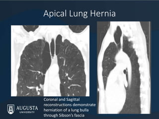
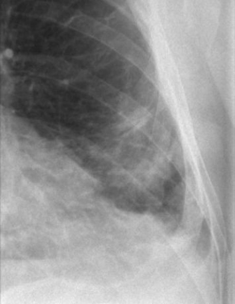
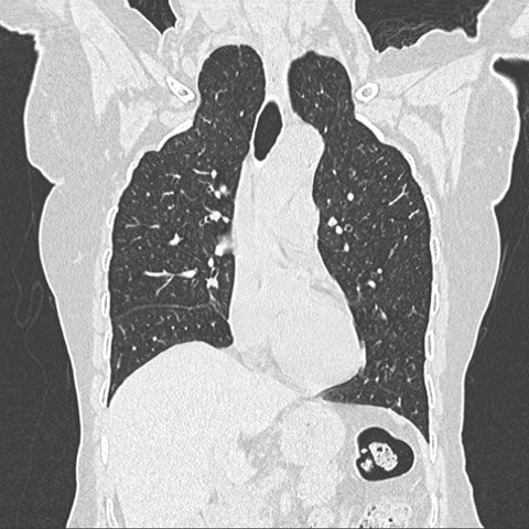
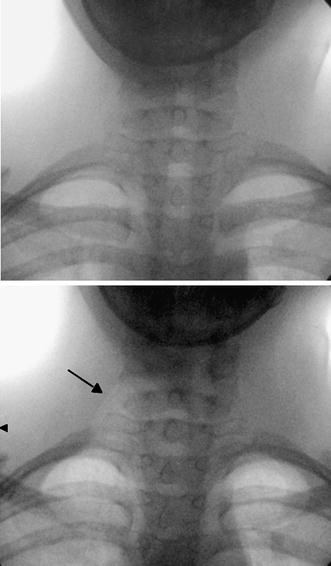
Apical lung hernias typically manifest as unilateral right-sided air radiolucencies at the thoracic inlet on chest radiographs. He was an ex-smoker with a. Airway fluoroscopy or CT pe.
Apical lung hernias typically manifest as unilateral right-sided air radiolucencies at the thoracic inlet on chest radiographs and can cause lateral tracheal deviation. OBJECTIVE We performed this study to characterize the clinical and radiologic manifestations of apical lung hernias. He had a 1-year history of a dry cough.
Institutional and Research4Life access to the MJA is now provided through Wiley Online Library. Dysphagia esophageal or coughing trachea 2. Surgical repair was not necessary as the hernias were asymptomatic and not associated with chronic cough.
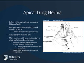
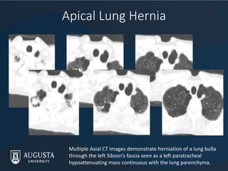
Apical lung hernias typically manifest as unilateral right-sided air radi- olucencies at the thoracic inlet on chest radiographs. This condition as a natural congenital occurrence is rareless than one in five cases reported. Herniation occurs through a defect in the Sibsons fascia and the apical segment of the lung protrudes in between the scalenus anterior and sternocleidomastoid muscles.
Apical lung hernias are often asymptomatic 1-3. Radiologic studies performed at midinspiration may not show hernias. The term pathological hernia is used to describe lung herniation induced by conditions such as tuberculosis focal bacterial infections empyema or osteomyelitis and malignant diseases 1.
Lung lobe herniation is the protrusion of lung parenchyma beyond the musculoskeletal thorax 14. 1 A hernia involving the cervical space has also been described as apical in the literature. Radiologic studies performed at midinspiration may not.
4 articles PMID. We performed this study to characterize the clinical and radiologic manifestations of apical lung hernias. Sometimes the diagnosis can only be made with a Valsalva maneuver which accentuates the herniation improving its visibility on physical examination.
Most people who experience a lung hernia suffered a severe trauma such as a traffic accident in which. Spontaneous apical lung herniation presenting as a neck lump in a patient with Ehlers-Danlos syndrome Ehlers-Danlos syndrome EDS is a heritable group of disorders of connective tissue characterised by skin hyperlaxity joint hypermobility and tissue fragility. The condition may be differed for the dog as spontaneous lung hernias were the most common reported as a sequelae of chronic respiratory diseases et al Choi 2015.
In humans lung lobe hernias are classified by location as cervical thoracic or diaphragmatic 14. In rare instances a lung hernia may become strangulated. A lung hernia refers to part of a lung pushing through a tear or bulging through a weak spot in the chest wall neck passageway or diaphragm.
A 50-year-old man was referred to us complaining of a swelling in his neck while coughing. The full article is accessible to AMA members and paid subscribers. 1Respiratory Medicine BYL Nair Hospital and Topiwala National Medical College Mumbai Maharashtra India.
About half of all lung hernias however appear after trauma to the chest 5. Some hernias of the lung however are symptomatic being accompanied by local pain paroxysmal coughing hemoptysis or any combination of the three. CONCLUSION Apical lung hernias typically manifest as unilateral right-sided air radiolucencies.
Apical lung herniation in adults is rare particularly in the absence of penetrating lung injury or chest wall disease1 2 It is due to a defect in the suprapleural membrane Sibsons fascia and small incidental apical parietal pleural defects have been described which may be present prior to the development of a larger defect3 4 Tearing of the fascia and spontaneous. Apical lung hernia. To the Editor.
It has rarely been reported in dogs 57. Symptoms when reported tend to be due to extrinsic pressure from the hernia on neck structures eg. Spontaneous apical lung herniation presenting as a neck lump in a patient with Ehlers-Danlos syndrome.
17874988 Indexed for MEDLINE Publication Types. Pulmonary hernias have been described through the diaphragm intercostal spaces and into the cervical space. This lung herniation was probably caused by a congenital deficiency in the suprapleural membrane Sibsons fascia combined with increased thoracic pressure created by the respiratory tract infection.
Overall 85 of lung herniation in humans was caused by trauma Moncada et al 1996. Apical lung hernias are often asymptomatic 1-3. Pulmonary herniation is an extension of the lung and pleura beyond their native positions in the thoracic cavity.
Symptoms when reported tend to be due to extrinsic pressure from the hernia on neck structures eg. They are frequently intermittent and can cause lateral tracheal. Apical lung hernia is a rare variety and has been confined to few case reports and series.
They are frequently intermittent and can cause lateral tracheal deviation. Surgeon 3149-51 01 Feb 2005 Cited by. Evans AS Nassif RG Ah-See KW.
Login to read more or purchase a subscription now. Apical lung hernias typically manifest as unilateral right-sided air radiolucencies at the thoracic inlet on chest radiographs.

Pdf Apical Lung Hernia Radiologic Findings In Six Cases
Intercostal Lung Hernia Radiology Case Radiopaedia Org
Apical Lung Hernia The Medical Journal Of Australia
2

Lung Hernia Radiology Reference Article Radiopaedia Org
2
Interesting Cases Of Lung Hernias
2

Bilateral Cervical Lung Hernia With T1 Nerve Compression The Annals Of Thoracic Surgery

Article Medicale Tunisie Article Medicale

Pdf Adult Cervical Lung Herniation Importance Of Valsalva Manoeuvre In Imaging
Dynamic Cervical Lung Herniation In A 10 Year Old Girl With Cough Springerlink

Figure 1 From Adult Cervical Lung Herniation Importance Of Valsalva Manoeuvre In Imaging Semantic Scholar

Pdf Adult Cervical Lung Herniation Importance Of Valsalva Manoeuvre In Imaging Semantic Scholar
Interesting Cases Of Lung Hernias
Thoracoscopic Repair Of Cervical Lung Hernia Sciencedirect
2
2

Apical Lung Herniation Radiology Case Radiopaedia Org
0 Response to "apical lung hernia"
Post a Comment